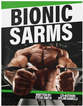
Bionic SARMS eBook
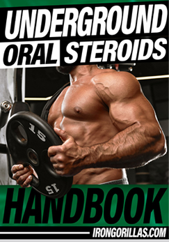
Underground Oral Steroid Handbook
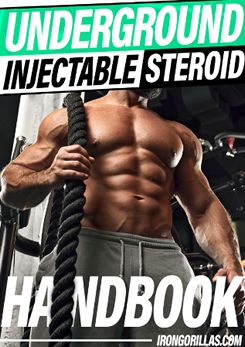
Underground Injectable Steroids Handbook
Anabolic Steroid Forums
Search
Featured Books
Bionic SARMS
Underground Oral Steroid Handbook
Underground Injectable Steroids Handbook
modal-check
“Bionic SARMs”: Your Ultimate Guide.
Download Now!
Email:
We respect your
email privacy
Dismiss ad
Dismiss ad
This will close in
120
seconds
modal-check
Underground Oral Steroid Handbook
Download Now!
Email:
We respect your
email privacy
Dismiss ad
Dismiss ad
This will close in
120
seconds
modal-check
Underground Injectable Steroids Handbook
Download Now!
Email:
We respect your
email privacy
Dismiss ad
Dismiss ad
This will close in
120
seconds